Image:Diatoms.png
From Wikipedia, the free encyclopedia


Size of this preview: 734 × 600 pixel
Image in higher resolution (1400 × 1144 pixel, file size: 951 KB, MIME type: image/png)
| | This is a file from the Wikimedia Commons. The description on its description page there is shown below. |
| Description |
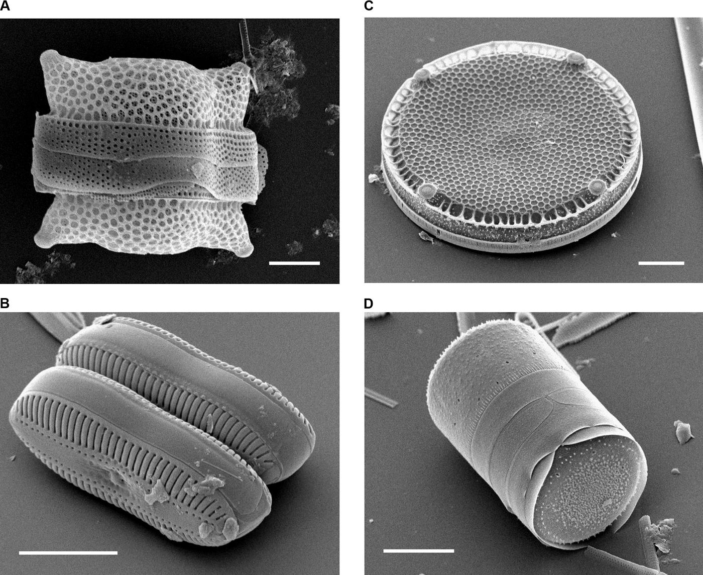
Scanning Electron Micrographs of Diatoms. (A) Biddulphia reticulata. The whole shell or frustule of a centric diatom showing valves and girdle bands (size bar = 10 micrometres). (B) Diploneis sp. This picture shows two whole pennate diatom frustules in which raphes or slits, valves, and girdle bands can be seen (size bar = 10 micrometres). (C) Eupodiscus radiatus. View of a single valve of a centric diatom (size bar = 20 micrometres) (D) Melosira varians. The frustule of a centric diatom, showing both valves and some girdle bands (size bar = 10 micrometres). |
||
|---|---|---|---|
| Source |
Bradbury J: Nature's Nanotechnologists: Unveiling the Secrets of Diatoms. PLoS Biol 2/10/2004: e306. http://dx.doi.org/10.1371/journal.pbio.0020306 |
||
| Date |
Published: October 12, 2004 |
||
| Author |
(Images courtesy of Mary Ann Tiffany, San Diego State University.) |
||
| Permission |
|

