Afbeelding:Amyg.png
Van Wikipedia

Amyg.png (189 × 230 pixels, bestandsgrootte: 22 KB, MIME-type: image/png)
| | Dit is een bestand van Wikimedia Commons. Onderstaande beschrijving komt van de beschrijving van het bestand daar. Controleer het gebruik van dit bestand in andere Wikimediaprojecten. |
| Description |
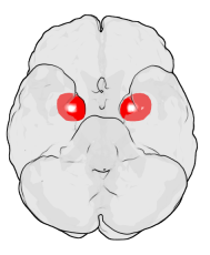
Location of the Amygdala in the Human Brain The figure shows the underside (ventral view) of a semi-transparent human brain, with the front of the brain at the top. The red blobs show the approximate location of the en:amygdala in the en:temporal lobes of the human brain. Note: the amygdala is covered by the ventral temporal cortex (i.e., it is inside the transparent brain). The figure was generated using en:MATLAB and Blender. It is based on MRI imaging data from the Wellcome Department of Imaging Neuroscience, UCL and on amygdalar coordinates from the Talairach brain atlas. If you like it, write on my talk page (en:User_talk:Washington_irving). I may be able to add some other brain regions. |
|---|---|
| Source | |
| Date | |
| Author |
User Washington irving on en.wikipedia |
| Permission |
Released under the GNU Free Documentation License. |
| Other versions |
Originally from en.wikipedia; description page is (was) here * 14:35, 13 February 2004 [[:en:User:Washington irving|Washington irving]] 189×230 (22,159 bytes) <span class="comment">(Location of the Amygdala in the Human Brain)</span> |

(Uploaded using CommonsHelper or PushForCommons)
Bestandsverwijzingen
Dit bestand wordt op de volgende pagina's gebruikt:

